

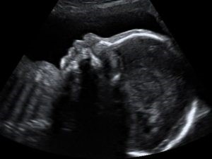
Here is one of the 4D baby images which I took during the routine ultrasound examination of the baby.
As you can see, the images taken during routine ultrasound are 2-dimensional, even if the parents understand this photograph during the examination, when viewed again later, the family often cannot understand which part of the baby this photograph belongs to.

With the 4-dimensional ultrasound technology, which has been used in recent years, we can obtain very high quality baby images, especially after the 26th week.
We give these images to our patients on CD or as photographs. Thus, when the family looks at it later, they can easily understand exactly which part of the baby has been watched.
The medical benefit of 4-dimensional ultrasound is that it provides the physician with more valuable information than routine ultrasound in terms of diagnosis of facial and skin anomalies in the baby.
Please click here to make a four-dimensional ultrasound appointment.